Deric Bownds
Upstairs/Downstairs in our Brain - What's running our show?
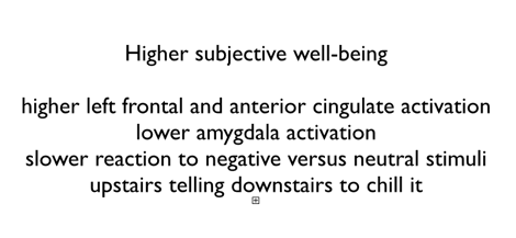
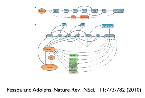
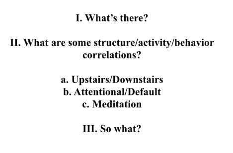
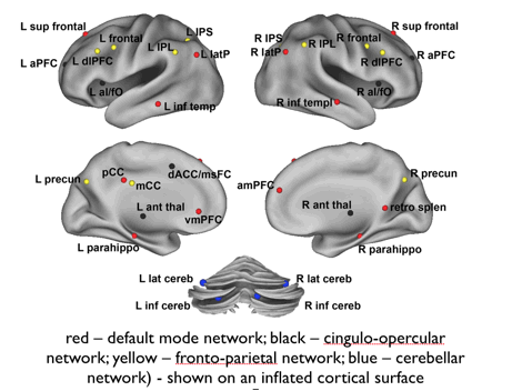
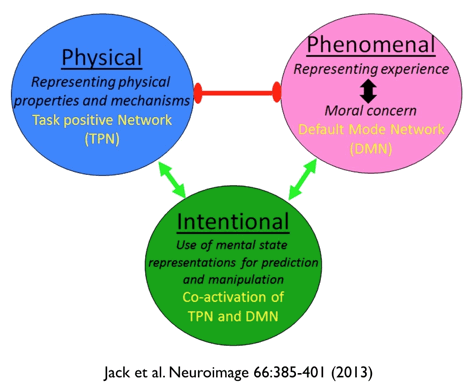
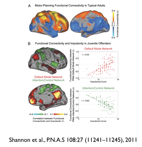
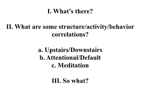
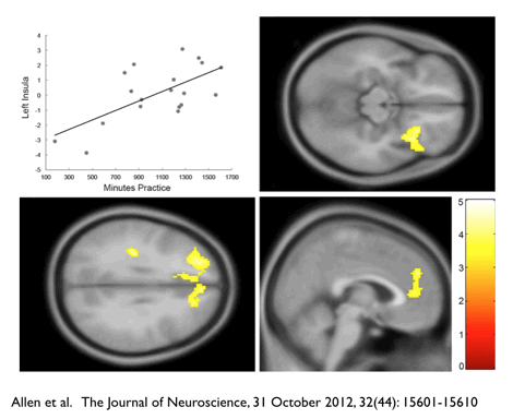
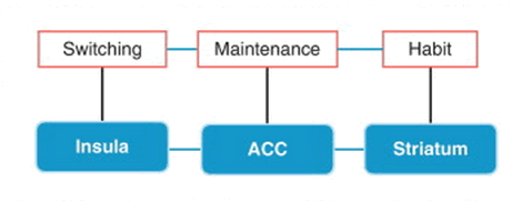
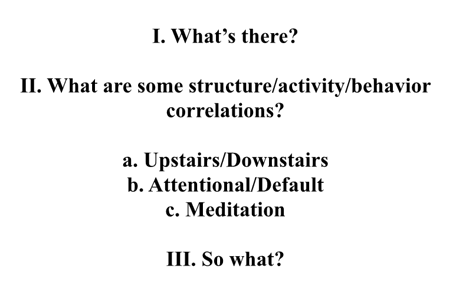
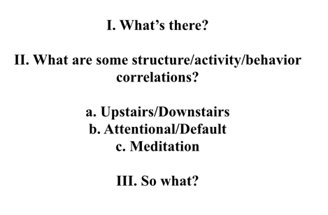
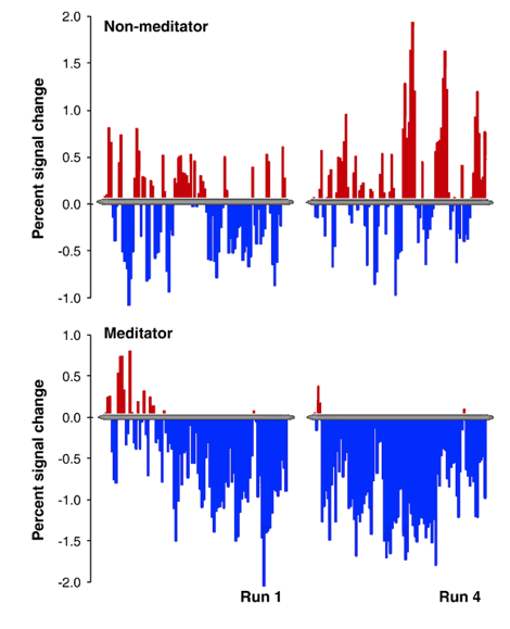
A talk given at the Sept. 9, 2014, session of the University of Wisconsin-Madison Chaos Seminar Series. Talk Abstract: Upstairs/Downstairs in our brains - What�s running our show? This talk starts with a brief brain 101 elementary anatomy review, and then offers a cherry picking review of recent trends in brain systems research that correlate what is going on in our brains with our behaviors. We want to know what normally makes us tick, what distortions might underlie addictive, impulsive, aggressive, stressed, depressed, or anxious behaviors, and what therapies might counter these distortions. I will focus on structure-activity-behavior correlations in three brain state distinctions that are currently being emphasized: Upstairs/downstairs and attentional/default mode systems that are a spontaneous part of our normal behavioral repertoire, and the cognitive therapy or mediation systems whose training, development, and expression can alter them. Lecture Text: Brains are a good topic for a chaos and complexity seminar. My first college biology course over 50 years ago had the standard joke �the brain has 1010 neurons, except for the cerebellum, which has 1012 and a common number you see is that the average neuron has 10,000 to 100,000 connections, each of which can be extremely complex and plastic, changeable. Those estimates have gone up since then, Current estimates are that a complete anatomical diagram would require at least one zetabyte, or 1021, or strictly, 270 bytes of information. This talk presents some brain 101 elementary anatomy and then notes some recent trends in brain systems research that try to relate what is going on in our brains to our behaviors. We want to know what makes us tick, and also perhaps tweak the system a bit when we are not getting things together so well. We would like to obtain a better understanding of what might be going on in OCD, stress, chronic depression, impulsive or aggressive behaviors. You frequently see descriptions emphasizing alternative brain states in a nice either/or bipolar way. Terms like Upstairs/Downstairs Top Brain/Bottom Brain System 1 (fast) /System 2 (slow) Conscious/Unconscious Attentional/Default Networks This can be partly correct, useful, but is a bit simple minded. We�re really dealing with complex networks with many nodes or hubs, like an air traffic network. I thought of switching the title to simply �what�s going on in our brains� but that didn�t sound very sexy, so I stayed with the upstairs/downstairs motif. There�s a lot of buzz today about brain science, Obama�s Brain Initiative, etc., so I thought I would start us off with a bit of showbiz that reinforces this and promises more than we can really deliver. We�ve come a long way from the crude PET images of Posner and Raichle that were getting us excited 20-25 years ago, you�ve probably seen these images, reproduced many times. The same areas that register more activity during vision, hearing, speaking are where strokes or other lesions compromise those function. These images are a far cry from all the bells and whistles of this 3-D animation of brain activity from Gazaley�s fly through brain. (You should be able to click on the image and view the video.)
This is supposed to be an anatomically-realistic 3D brain visualization depicting real-time source-localized activity (power and "effective" connectivity) from EEG (electroencephalographic) signals. Each color represents source power and connectivity in a different frequency band (theta, alpha, beta, gamma) and the golden lines are white matter anatomical fiber tracts. Estimated information transfer between brain regions is visualized as pulses of light flowing along the fiber tracts connecting the regions. Here is the descriptive gobbledegook (to me, that is) that goes with the movie� please let me know if you find a refereed scientific article that has this information, as of August 2014 I haven�t been able to. Carl Zimmer's article that pointed me to this said that "the volunteer was simply asked to open and shut her eyes and open and close her hand." If so, what about at least labeling the moving graphics "eye shutting" "eye opening" "hand opening" "hand closing," and could they maybe tell us which colors refer to which frequency bands? Very frustrating. I dug around for a while on their websites I could find anything approaching a manuscript or reference information, but wasn�t willing to spend more time on it. This is an extreme example of the kind of presentation people doing psychology and cognitive neuroscience seem to feel compelled to offer these days, whether due to the difficulty in attracting funding or the quest for celebrity status. You can�t just send in a paper and have it reviewed and published. You have to do a press release through your institution, fire off an op-ed piece to the new york times, get your agent to send a spiel to science writers and bloggers, maybe even before the work is submitted for publication, and certainly by the time it under review or accepted for a forthcoming date. At least once a week I get a �we think your MindBlog readers would be interested in�. fill in the blank.) I�ll show you another gee-whiz macro depiction that comes from the new Clarity technique, which uses a clever mixture of solvents to replace nerve membrane lipids with a transparent gel so that fluorescently labeled neurons can be visualized. Here�s a 3-D animation of a mouse brain�s neurons (Again, click to play).
I�ll throw in a final visual treat that gets down to the ultrastructure of individual nerve cells. Getting into detailed brain wiring requires the analysis of thousands of serial sections of electron microscope images, here is a reconstruction of a group of retinal bipolar cells. This work is extremely laborious �50 hrs to do one retinal neuron from electron microscope serial sections. Seung, at MIT and now Princeton, has asked for help from the general public (�citizen neuroscientists�) to crowd source it. Their data on how bipolar cells connect to amacrine motion detecting cells in the retina has suggested a model for motion detection (two bipolars, first delays output so arrival at amacrine is simultaneous, this might signal motion of moving spot that serially excites the bipolars.) This model is hand waving until electrical recording are done from the suggested circuit�s cells. This animation of their results at the indicated URL is a treat to watch. (Click to watch).
Knowing this anatomy doesn�t get you there, a functional description would have to include information that synapses are awash in neurotransmitters, hormones, modulatory peptide (small proteins), neural-growth factors that alter the transmission of signals. Synapses constantly form and dissolve, weaken and strengthen, in response to new experiences. Old brain cells die, new ones are born, genes are constantly turning on and off. The brain is most likely processing information at many levels below and above that of individual neurons and synapses�. Obama�s Brain initiative and the Brain Activity Map project can only be groping to define the playing field, rather than assuming that we now know what it is. As noted in the above summary graphic by Grillner, we�re not going to be able to avoid detailed and tedious studies of the many levels of organization that go into building a behavior. OK, enough of the fancy graphics. So, three questions make the gestalt for this talk: a general introduction on what�s there, then get into some structure-activity-behavior correlations focusing on these three brain state distinctions that recent work has more explicitly distinguished: Upstairs/Downstairs, attentional/default, meditation systems. And then a bit on the �so what?� question. Why do I list meditation? It is not like the Upstairs/Downstairs and Attentional/Default mode networks that are spontaneously going on all the time - it requires training. Well, because meditation or cognitive therapy correlates with brain changes and is training in discriminating where we are at - at a given moment - with respect to IIa. and IIb., and perhaps allows us to have some influence on their expression. I�ll start with a bit of brain 101, reminding of some core areas it is useful to be familiar with. The assignment of functions to brain areas has come from correlating sensory or motor behaviors with imaging or electrical recordings, as well as noting the behavioral effects of regional brain damage from strokes or tumors. The figure labels the primary visual, auditory, somatosensory, and other areas. In general information coming in first projects to the back of the brain and then moves forward in a dorsal stream that is figuring out the spatial relationships in the environment so that it can act on them and a ventral stream that is concern with the identity and meaning of what is coming in. The frontal lobes that are focusing attention actually feed back to the primary sensory areas to enhance processing of what�s most relevant. Kosslyn and Miller have done a recent trade book called �Top Brain - Bottom Brain: Surprising In sights into How You Think.� in which they offer this ventral/dorsal pathway distinction as a general description of a bottom brain system that classifies and interprets information from the world and a top brain system that formulates and executes plans. They charge on to explain four basic personality types based on this simple distinction: The Movers are your winners, top brain action people who actually also use the bottom have to pay attention to the consequences of their actions and use the feedback. Their stimulator is more a �damn the cannons, full speed ahead� kind of person, less inclined to attend to bottom pathway, the consequences of their actions and know when enough is enough. The Perceiver is mainly bottom brain interpreters, unlikely to initiate top brain detailed or complex plans. Finally, the people with lazy top and bottom brains are your �whatever�� types, absorbed by local events and immediate imperative, responsive to ongoing situations. i.e. the U.S. electorate. Other popular books offer alternative modern phrenologies that emphasize different sets of functions. The most common drawings you see, like this, just show the external surface of the neocortex, We need to look also medial views like the right side of this figure of divisions of the frontal lob. and also cross sections showing how the convoluted cortex curls around on the inside: I�ve labeled cingulate, amygdala, insula - all considered part of the inside or bottom brain limbic lobe system. Here is a slide of a cross section with colors marking the areas. The insula is like the sensory cortex of interior self, sensing viscera, interoceptive awareness, homeostasis, emotions like disgust, social emotions, part of the downstairs brain limbic system. The amygdala has a primary role in emotional reactions, also decision making and memory. The right amydala activity correlates more with negative stuff and left amygdala activity with positive stuff. The cingulate cortex lies just over the corpus callosum, the bundle of fibers linking the two hemispheres, sort of linking cognitive frontal functions with emotions. Error detection, conflict monitoring, pain, motivation and reward. The insula plus the dorsal anterior cingulate cortex connect as the central part of the salience network that gives the first cognitive signal of behaviorally salient events such as errors. These internal structures are harder to nail down than the outer layers of the cortex (visual, motor, somatosensory, auditory, etc.), have their fingers in many homeostatic and other functions, (If you lesion the former, you get fairly tidy agnosias of vision, hearing, or smell, or motor deficits of some sort, lesions of the latter generate more chaotic global effects). More recent work has moved beyond the above visualization of large blobs of areas to get down to finer parcellations, down to about 1000 regions of interest, arrived at by using a flavor of MRI, magnetic resonance imaging, called diffusion tensor imaging that looks at perturbations of water diffusion caused by obstacles, from which the paths of bundles of axons, i.e. white matter tracts, and brain connectivity (bottom right of figure below) can be computed. The connectivity profile of brain networks looks something like an air traffic network with larger and smaller airports� a group of 12 strongly interconnected bihemispheric hub regions form, called a �rich club� comprised of frontal and parietal cortex, precuneus, cingulate and the insula, as well as the hippocampus, thalamus, and putamen. This is work published in 2011. More recent work has done a more fine grained analysis finding subnetworks, and suggesting alternative hubs. You can watch these connectivity networks go bonkers during something like watching a scary movie when your pumping adrenaline, the blue stuff shows brain regions that get more active and connected, averaged over many subjects. β-adrenergic receptor blockade diminishes this increase. Exposure to acute stress prompts a reallocation of resources to a salience network, promoting fear and vigilance, in which the dorsal anterior cingulate and anterior insulas are central at the cost of an executive control network. After stress subsides, resource allocation to these two networks reverses, which normalizes emotional reactivity and enhances higher-order cognitive processes important for long-term survival. Nudging more into the second topic, II activity behavior correlations, Let�s start with upstairs/downstairs. Here is an example of the sort of graphic you can see in the literature: This is from work of Kevin Ochsner and colleagues at Columbia who used functional magnetic resonance imaging to compare the neural correlates of negative emotions generated by the bottom-up perception of aversive images and by the top-down interpretation of neutral images as aversive. They found that both types of responses activated the amygdala, although bottom-up responses did so more strongly - bottom-up responses activated systems for attending to and encoding perceptual and affective stimulus properties, whereas top-down responses activated prefrontal regions that represent high-level cognitive interpretations -self-reported affect correlated with activity in the amygdala during bottom-up responding and with activity in the medial prefrontal cortex during top-down responding. Looking at data like this makes descriptions like top brain/bottom brain, upstairs/downstairs seem a rather crude way of putting it�you�re seeing activities all over the place, but the blue stuff, that shows greater activity for top-down processing, does in general reside above the yellow orange stuff, greater for bottom up than top down. Here is another illustration of downstairs and upstairs response systems from Dolan�s lab at the Welcome Inst. in London. Here they are watching people play a computer game in which they are in a maze, and are told that they are being pursued by a virtual predator that can chase, capture, and inflict pain. When the predator is perceived as being at some distance moving in the maze is more a cognitive escape puzzle, but as the virtual predator draws close, things get a bit more gutsy, and brain activity shifts from the ventromedial prefrontal cortex to the periaqueductal gray, along with an increased subjective feeling of dread and decreased confidence in escape Another example�Etkin and his collaborators at Stanford have done a meta analysis of a large number of studies showing that in anxious and depressed individuals the amygdala, insula and superior temporal gyrus, and dorsal anterior cingulate are over-reactive while the Dorsolateral prefrontal cortex and caudate body are under-reactive. (i.e. the brain�s downstairs is over-riding the upstairs.) Cognitive training exercises that reinforce a positivity bias and enhance working memory (they are easily available on the web) lessen this upstairs/downstairs imbalance. And, MRI data indicates an enhancement of medial prefrontal activity. So, this is an example of upstairs telling downstairs to chill, and here is another one: Kober et. al. do a variation of the classic you can have one piece of candy right now or two if you wait awhile. Later versus Now trials. The top frame shows medial and lateral views of brain regions that show greater activation in the LATER vs. NOW trials, when study participants use a cognitive strategy to reduce their craving. The circled or highlighted activations are in regions know to be involved in regulation of aversive emotion. The bottom frame shows coronal and medial views of brain regions that have reduced activation in LATER vs. NOW trials. The circled highlighted reductions are in regions involved in cue-induced craving or emotion. [corr, Corrected for multiple comparisons; uncorr, uncorrected for multiple comparisons.] In general high subjective well being correlates with left frontal activation and anterior cingulate activation, lower amygdala activation, the upstairs telling the downstairs to chill. This figure from my book describes another feature of the upstairs/downstairs motif. Downstairs is fast, upstairs is slow. Downstairs offers a quick and dirty route that bypasses higher cognitive perception and interpretation to get the amygdala excited and initiating avoidance movement before higher centers have decided whether the shape in your peripheral vision is a snake or a wavy piece of rope on the ground. Of course it couldn�t be that simple, subsequent work has shown that there are multiple waves of activation in pathways from retina to amygdala that involve feedback. In terms of behavior correlations, these examples have been contrasting cognitive frontal mainly inhibitory controls than can regulate more rapid medial lower areas like the amygdala where habitual routines and learned emotions reside. It takes increased activity in our frontal cortex to override addictions and cravings fueled by deeper structures in our old mammalian brains, or to dampen the inappropriate reactivity of our amygdala. Ideally there is an upstairs downstairs balance that does not allow either uninhibited limbic emotional reactivity or a frontal control freak to run the show. Distinguishing upstairs more frontal and cognitive areas from downstairs more medial emotional regions segues into another important distinction, the attentional versus default systems. Here are the kinds of descriptions you see in the literature: I�m going to zoom through a number of summary depictions of these systems so you can get the general gestalt, you�re probably not going to want to get immersed in trying to remember all the regions or anatomy. The attentional and control system is more frontal and lateral - The blue stuff in this graphic from a Buckner review article The default system. the red and yellow stuff, is associated with the medial temporal and frontal inside of the brain, that participates in internal modes of cognition, when we are not focusing on external environment, task oriented. Here is what lights up during different kinds of internal mentation�thinking about future, theory of mind, etc. These default regions are only sparsely functionally connected at early school age (7–9 years old); over development, these regions integrate into a cohesive, interconnected network. Here is a slightly more fine grained breakdown of nodes in these systems from Fair et al. (PLOS Computational Biology (DOI: 10.1371/journal.pcbi.1000381): This slide color codes points, hubs, or nodes in four main networks that can be distinguished in the attentional and default systems. Red is the default mode network - bilateral posterior cingulate/precuneus, inferior parietal cortex, and ventromedial prefrontal cortex - all show a decrease in activity during goal-directed tasks. Yellow and black get more active in goal-directed tasks, yellow a frontal parietal network that acts on shorter timescales to initiate and adjust top-down control. The black cingulo-opercular network operates on a longer timescale providing �set-initiation� and stable �set-maintenance� for the duration of task blocks. Along with these two task control networks, a set of blue cerebellar regions showing error-related activity across tasks forms a separate cerebellar network that connects to both the yellow fronto-parietal and black cingulo-opercular networks. The networks develop from a local to distributed organization during development. Here is the final visualization of task positive versus default mode activities that I will flash up, done by Jack et al. Here is their MRI imaging summary of brain regions whose activity correlates with these distinctions in both hemispheres, left is lateral, center medial, and right is both graphically splayed out as a continuous sheet. Blue and green are physical task positive areas, mostly outside lateral parietal frontal surface of cortex, yellow and red are default mode experiential social mostly inside medial, Their fMRI measurements show reciprocal inhibition between social and physical cognitive domains. Empathy and social cognition suppress analytic thought, and vice versa. We have a physiological constraint on our ability to simultaneously engage these two distinct cognitive modes. They suggest that where their activities overlap could be intentional use of mental state representations for prediction and manipulation, a co-activation of task positive and default mode systems. Here is their cartoon summary of the specializations of attentional and default mode and their collaboration. Just as with our upstairs/downstairs behavior drivers there is an optimal balance between attentional and default modes. With respect to their behavioral correlates, they both get a good press and a bad press. In general the attentional mode is valued as giving our control to more frontal cognitively and metabolically demanding activities. It is needed for episodic memory formation, along with deactivation of the default mode network. Its downside can come with being a control freak, too much attentional control, suppression of spontaneity and creativity. The attentional upstairs frontal stuff is more susceptible to aging, and most of us find it more of an effort to do novel versus habitual actions as we age. A default mode wandering mind has its virtues and constructive aspects: self- awareness, creative incubation, improvisation and evaluation, memory consolidation, autobiographical planning, goal driven thought, future planning, retrieval of deeply personal memories. Simple experiments demonstrate how mind wandering can facilitate creative incubation, that the basement stuff that is dinking around in the absence of focused effort more easily makes novel associations. The bad press for the default mode includes a clever study by Killingsworth and Gilbert titled �A wandering mind is an unhappy mind.� (They recruited thousands of people to use a cell phone App to note their mood when prompted and also say what they were doing at the time.) There is a consensus that excessive rumination or mind-wandering, activity in the default mode network may predispose people to depression, anxiety, attention deficit, and post-traumatic stress disorder. Increasing activity of the default mode network is associated with passive reactivity and using environmental support rather than focused frontal self-initiation. Environmental support has a bright and a dark side: it helps aging individuals to perform but comes with a loss in internal control. I wonder if an excess of default mode activity might be at issue in the diagnosis du jour, especially in children, called sluggish cognitive tempo (i.e. sluggish attentional mode), that is pushing aside ADHD. Maybe this is characteristic of a brain that is chronically in the default mode and unable to (or unwilling or too lazy to) activate the �attentional� or goal oriented, direct experiential focus, task positive network appropriately. (Think about the teenagers behind fast-food counters completely unable to remember do simple addition and subtraction, or remember what you just told them about to go or for here, or soft shell or hard shell taco!). Another point is that impulsive or delinquent behaviors appear to correlate with more than average default mode and less attentional mode activity. The above is a graphic from Shannon et al, who examined resting-state functional connectivity among brain systems and behavioral measures of impulsivity in 107 juveniles incarcerated in a high-security facility. In less-impulsive juveniles and normal controls, motor planning regions were correlated with brain networks associated with spatial attention and executive control. In more-impulsive juveniles, these same regions correlated with the default-mode network, a constellation of brain areas associated with spontaneous, unconstrained, self-referential cognition. The interest of groups studying the attentional/default and upstairs downstairs systems is now overlapping with those that have been working on behavioral and brain correlates of meditation. So lets move on to that for a bit. Meditation is not something that �just happens� like upstairs versus downstairs, attentional versus default activities. It requires training and practice, in blocks of time set apart from normal daily activities, and so the emphasis has been on figuring out whether long term changes in the brains of meditators can be seen that correlate with changed behaviors. The problem is that �Meditation practice� can refer to a wide range of training regimes, some directed at strengthening the attentional mode and others, the default mode. Mindfulness meditation can refer to various training strategies that emphasize or include attention regulation, body awareness, emotion regulation, or changing self-perspective. In most mindfulness practices and other techniques linked to Buddhist traditions, mind wandering is considered a distraction and a gateway to rumination, anxiety and depression, so a goal is to reduce it in favor of attentional mode. Other practices go for spontaneous non-directive inner mind wandering, Transcendental Meditation, Benson�s Relaxation Response, Clinically Standardized Meditation, Integrative Body-Mind regimes (IBMT) regimes, also originating from Eastern traditions, make no effort to control thoughts, but instead a state of restful alertness that allows a high degree of awareness of the body, breathing, and external instructions. Brain changes are seen in practitioners of these strategies, although most of the experiments are not rigorous enough to attribute brain changes to mindfulness practice per se rather than to pre-existing differences in the experimental and control groups. Grazing the literature I find several papers saying mindfulness training increases resting state or default activity and several others saying it decreases it. The simple fact is that any specialized training, of either a skilled physical or introspective ability, causes long lasting brain activity and/or structural changes (such as increases in gray matter volume) that persist as long as the activity in question is practiced and maintained, and that return to the average if it is discontinued. The brain�s representation of the hands is enlarged in skilled pianists. It�s as simple as �Use it or lose it�. Richie Davidson and his collaborators have a study suggesting that a kind of meditation I haven�t mentioned yet, compassion training, increases activation of brain areas involved in mirroring, empathy and social cognition. If you go zoomin� around the web you�ll find reports of MRI data showing larger or smaller Gray Matter volumes here, there, and the next place in meditators versus controls. Mindfulness based stress reduction correlates with decrease in size of amygdala. A number of studies are short term, the behavioral and brain changes after a six weeks training regime are measured and compared with controls In the above study by Allen at al., mindfulness training increased dorsolateral prefrontal (bottom left), right anterior insula (top right), and medial–prefrontal BOLD (bottom right) recruitment during negative emotional processing. This is after only six weeks of training On balance it seems that expert meditators, of either focused attention, open monitoring, compassion styles show deactivation of the main nodes of the default-mode network. The above work from Brewer et al. on expert meditators shows coactivation of mPFC, insula, and temporal lobes during meditation. These people are able to control cognitive engagement in conscious processing of sensory-related, thought and emotion contents, by massive self-regulation of fronto-parietal and insular areas in the left hemisphere The insula appears prominently in many reports: The above study by Luders et al. comparing 22 controls with 22 people experienced with at least five years of various forms of meditation. They found significantly larger gyrification and gray matter volumes in meditators in the insula regions involved in emotional regulation and response control. Larger volumes in these regions might have something to do with meditator�s abilities and habits to cultivate positive emotions, retain emotional stability, and engage in mindful behavior, to integrate autonomic, affective, and cognitive processes. Posner et al have done a summary of brain correlates of default resting, attentional alert, and meditation states. In this eyes-glaze-over summary table listing different brain areas and neurotransmitters associated with the three states, stage 1 refers to early effortful training that invokes lateral prefrontal and parietal cortex, stage 2 is flickering back and forth between attention and mind wandering, stage 3 is the advanced stage of training, usually thought to be obtained with little or no effort, meditation is maintained by activity in the ACC, left insula, and striatum, and connectivity between these regions is enhanced by one month of training. There is parasympathetic dominance with strong activation in the ACC and left insula, and reduction of activity in the lateral PFC and parietal cortex. Their model for the neural correlates of changing brain states includes the insula, ACC, and striatum (IAS). They suggest the ACC is involved in maintaining a state by reducing conflict with other states; the insula serves a primary role in switching between states; the striatum is linked to the reward experience and formation of habits required to make state maintenance easier. If we move on to the behavioral correlates of meditation (IIc), most studies being on mindfulness meditation, the literature mainly lists beneficent effects. There doesn�t seem to be much sentiment that you can have too much mindfulness, like you can have too much downstairs versus upstairs or default versus attentional mode activity. Now, back to continue with topic III the �so what� of the brain behavior correlations I been illustrating. We�ve done a bit of defining upstairs, downstairs, attentional, default, and meditation modes and their anatomical and behavioral correlates - in general external attention, focus, control emphasize frontal and parietal lateral areas and medial inside regions engage for the downstairs and default mode activity, with insula, ACC, and striatum choosing and holding in one or another state. We would like to use our awareness of what is going on to influence it in a useful and adaptive way, enhance behaviors that work and diminish those that don�t. We�ve known since Wilder Penfield�s work in the 1950s that our conscious awareness can influence the most intimate details of our nerve cells firings. He used the electrode that was being used to probe the brain regions about to undergo epileptic surgery to also record activity of single nerve cells, letting their firing be played on a loudspeaker as staccato bursts of noise. When he instructed the patients to slow down and try to stop those bursts, they could do it. There are thousands of published reports of using autonomic nervous system feedback, skin conductance, heart rate, blood pressure, or various kinds of e.e.g recordings or MRI recordings to alter alertness, calm, stress, etc. The figures below are from a Yale group looking at real-time fMRI feedback from the Posterior Cingulate Cortex, a hub of the default mode network shown to be activated during mind-wandering and deactivated during meditation. Both meditators and non-meditators reported significant correspondence between the feedback graph and their subjective experience of focused attention and mind-wandering. The arrow points from posterior cingulate to increased activity corresponding to subjective mind wandering, the blue is the decrease in activity during focused attention. When instructed to volitionally decrease the feedback graph, meditators, but not non-meditators, showed significant deactivation of the posterior cingulate cortex. There is a consensus that excessive rumination or mind-wandering, activity in the default mode network may predispose people to depression, anxiety, attention deficit, and post-traumatic stress disorder. So the use of meditation, brain exercises, or cognitive therapy techniques to enhance attentional network activity is therapeutic. It doesn�t require that we hook ourselves up to electrical widgets or brain imagers. How in fact might this work? We�ve seen MRI or other recordings of activity increases that correlate with various behaviors, where would we see activity if we were observing and changing our own brains, the ratio of attentional and defaults or upstairs and downstairs influence. Its known that temporal-parietal lobe junction (TPJ) and superior temporal sulcus (STS), are central to integrating and shifting attention, perspective taking and theory of mind, so these areas, whose volume is increased in meditators, are good candidates for part of that system. As I was working up the list of factoids and figures I�ve been throwing at you I kept wondering to myself, how do I wrap this kind of material up, draw us to a close? Does knowing this make us some kind of super person? No� but I guess it satisfies our curiosity to know something about ourselves, and it probably helps more than it hurts our individual trips to have some ability to distinguish what brain states are resident at a given moment in our ongoing opera. And, we can choose to engage brain exercises, or meditation techniques, whose practice lets us better control what state we are in. I would submit that those mind therapies, meditations, or exercises that are the most effective in generating new more functional behaviors are those that come close to resolving what we could call the category error (in the spirit of the philosophical term) in considering mind and brain. And, that error is to confuse a product with its source, the source being the fundamental impersonal downstairs machinery that generates the varieties of functional or dysfunctional selves that are its product, that we mistakenly imagine ourselves to be. Mental exercises like meditation permit the intuition of, perhaps come closest to, that more refined metacognitive underlying generative space that permits viewing of, and choice between, more or less functional self options. A less wordy, maybe more useful, way of putting this is to say that third person introspection, viewing yourself as if looking at another actor, and placing this a historical story line, is more useful than immersed rumination (coulda, shoulda, woulda). It is the difference between residing mainly in the attentional versus default modes of cognition. If there is a practical take-home message, it is that maintaining awareness of, and exercising, focused attentional mechanisms is important to mental vitality and longevity. Such awareness is central in resisting the attacks on our attentional competence that comes from the confusing media jungle that tempts our passive default mode receptivity and reactivity. |


